Original source: Medical Physics, Radiation Oncology & Cancer
With: Reinoud van der Straeten · Juliana Simoes
This article is an editorial summary and interpretation of that content. The ideas belong to the original authors; the selection and writing are by Streamed.News.
This video from Medical Physics, Radiation Oncology & Cancer covered a lot of ground. 6 segments stood out as worth your time. Everything below links directly to the timestamp in the original video.
Understanding this unique stopping power is crucial for appreciating why proton therapy can reduce side effects and improve outcomes for patients, especially those with tumors close to sensitive organs.
Protons Deliver Precise Dose to Tumors While Sparing Healthy Tissue
Proton therapy offers a significant advantage in cancer treatment due to its ability to precisely stop within tissue, a benefit highlighted by Reinoud van der Straeten, a product manager at Varian. Citing Tony Lomax, head of physics at PSI in Switzerland, van der Straeten explained that this allows clinicians to deliver 100% of the radiation dose directly to a tumor, such as one ending at a depth of 7 cm, while ensuring 0% of the dose reaches an organ at risk located immediately beyond the target area. This precision is achieved by carefully selecting proton energy, like 96 MeV, to ensure the Bragg peak—the point of maximum energy deposition—terminates exactly at the tumor's distal edge.
"The benefit of using protons is that they stop in the tissue."
Uncertainty in Proton Stopping Depth Poses Challenge for Therapy Planning
While protons offer precise dose delivery, a critical challenge in proton therapy is the inherent uncertainty in exactly where these particles will stop within tissue, according to Reinoud van der Straeten from Varian. Even a 3.5% range uncertainty can significantly shift the Bragg peak—the point where most radiation is deposited. This means a planned 7 cm stopping depth could, in reality, be shallower, causing the entire radiation dose to shift from the intended tumor target into a nearby organ at risk, potentially exposing it to 100% of the dose instead of 0%.
"This is what we need to mitigate when we are doing proton planning."
Eclipse Defines Patient and Range Uncertainties for Robust Proton Therapy Optimization
To achieve robust proton therapy plans, the Eclipse planning system allows users to define specific perturbations that account for real-world uncertainties. This includes modeling patient positioning errors, where the isocenter can shift by 3mm in the X, Y, and Z directions, generating six distinct scenarios (plus and minus for each axis). These shifts represent common variabilities in patient setup during treatment.
In addition to positional uncertainty, the system incorporates a 3.5% stopping power uncertainty for range, which means the effective tissue density for each voxel in the patient can vary by plus or minus 3.5%. This accounts for variations in how protons slow down in different tissues. Combining these factors results in eight different uncertainty scenarios, which are then used alongside the nominal plan to assign nine different dose calculations to each voxel during the robust optimization process.
"I need to tell the system what kind of uncertainties I have."
Eclipse Begins Robust Optimization, Visualizing Dose Uncertainty with Robustness Bands
Initiating robust optimization in Eclipse involves a multi-step computational process where the system first calculates beam lines and then defines the necessary energy layers and individual spots to cover the target volume. The software subsequently optimizes the weights of these spots, effectively determining the fluence map to deliver a homogeneous dose to the target area while considering all defined uncertainty scenarios.
During this optimization, the Dose Volume Histogram (DVH) displays critical "robustness bands" alongside the nominal dose line. These bands visually represent the range of minimum and maximum doses that each voxel within the target structure might receive across all perturbation scenarios. The primary goal of robust optimization is to narrow these bands as much as possible, ideally converging them into a single line, indicating that the target will consistently receive the same dose regardless of uncertainties.
"You also see these bands, what we call the robustness bands… The idea is to ensure that this band is acceptable."
Tissue Heterogeneities Dramatically Shift Proton Dose Distribution
Tissue heterogeneities, or variations in density within the body, significantly impact how proton beams deliver radiation. A demonstration showed that introducing a lung cavity into a homogeneous water phantom caused the proton dose distribution to visibly shift downwards, even when using the exact same treatment plan. This occurs because the lung tissue has a lower "water equivalent thickness"—around 11.8 cm compared to 13.6 cm for water in a specific region—meaning protons travel further through it.
This dramatic shift underscores the critical importance of obtaining precise density information for patient tissues in proton therapy planning. Without accurate data, the intended homogeneous dose coverage can be compromised, leading to underdosing the target and potentially overdosing healthy tissues beyond it. While heterogeneities pose a challenge, planning systems can account for them by using different beam energies for various parts of the target, ensuring a consistent dose distribution.
"You can see here how important the effect of a different density is."
Evaluating Robust Proton Therapy Plans Through Uncertainty Recalculation
After a proton therapy plan undergoes robust optimization, merely reviewing its nominal dose distribution is insufficient for a comprehensive evaluation. The next crucial step involves a robust evaluation, where the treatment plan's stability is rigorously tested against the same spectrum of uncertainties that were considered during its optimization. This ensures the plan remains effective under anticipated real-world conditions.
During this evaluation, the optimized plan is recalculated across all predefined uncertainty scenarios. This includes re-evaluating the dose distribution with simulated patient positioning shifts—specifically, 3mm displacements in the X, Y, and Z directions—and accounting for a plus or minus 3.5% variation in tissue stopping power, which affects the proton range. This systematic recalculation for each scenario allows clinicians to thoroughly assess the plan's resilience and confirm its robustness before treatment.
"We also need to be able to evaluate the plan in a robust way."
Also mentioned in this video
- The Bragg peak phenomenon in proton therapy, contrasting its significant dose… (1:07)
- Different proton delivery techniques, focusing on modulator scanning as the… (3:08)
- The presenter defines a proton 'spot' as a narrow, monoenergetic beam,… (3:43)
- The use of multiple superimposed energy layers or Bragg peaks to create a… (5:01)
- The presenter illustrates how modulator scanning works by using multiple energy… (5:47)
- The cyclotron and energy selection system in proton therapy, explaining how a… (7:23)
- The function of a range shifter, a polycarbonate plate inserted into the… (8:49)
- Target and uncertainties in proton planning, highlighting the main uncertainty… (10:54)
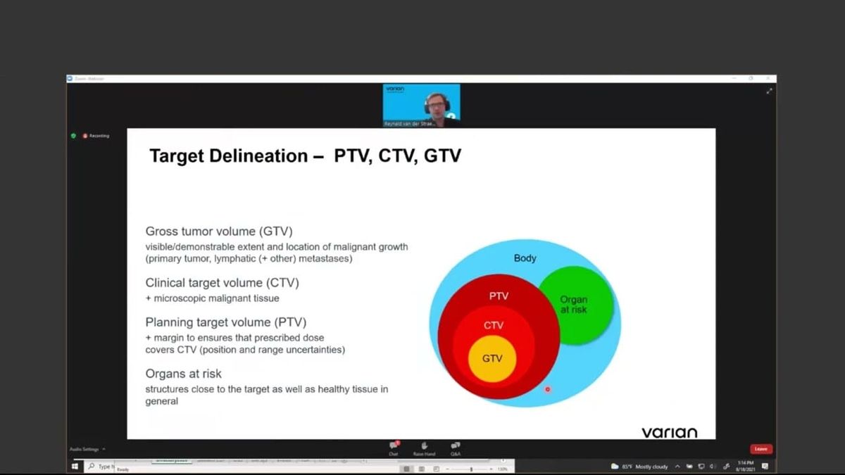
- Unlike photon therapy which uses a PTV, proton planning focuses on the CTV due… (11:51)
- The presenter provides a basic illustration of how proton planning considers… (13:09)
- The presenter concludes the theoretical discussion by reiterating that range… (16:27)
- The presenter transitions to a live demonstration in the Eclipse workstation,… (17:01)
- The presenter resumes the Eclipse demonstration, showing a single spot's Bragg… (18:43)
- The presenter demonstrates adding multiple energy layers in Eclipse to achieve… (20:32)
- The presenter demonstrates the use of a range shifter in Eclipse to enable the… (21:50)
- The presenter transitions to a head and neck case in Eclipse, explaining that… (26:23)
- The presenter demonstrates using RapidPlan in Eclipse to automatically generate… (27:06)
- The presenter demonstrates setting robust objectives in Eclipse for the CTV,… (29:55)
- The presenter demonstrates evaluating the robustness of the plan in Eclipse by… (36:24)
- The presenter concludes the demonstration of how to evaluate and optimize a… (39:55)
Summarised from Medical Physics, Radiation Oncology & Cancer · 44:45. All credit belongs to the original creators. Streamed.News summarises publicly available video content.
Streamed.News
This publication is generated automatically from YouTube.
Convert your full video library into a digital newspaper.
Get this for your newsroom →